


Screening tests often find breast problems, such as cancer, before there are warning signs or symptoms. Treatment is most successful when it starts at this early stage. This is why you should talk to your primary care or women’s health provider about how often you need to schedule regular screenings.
Our partners at Dartmouth Cancer Center provide more information about non-cancerous breast conditions and breast cancer.
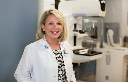
Cheshire Medical Center’s breast radiology and imaging team includes highly-skilled radiologists and mammography, ultrasound, and magnetic resonance imaging (MRI) technologists who provide our advanced diagnostic capabilities.
Radiologists interpret screening results and perform tissue biopsies using diagnostic imaging scans to guide the procedure. They work collaboratively with the pathologists, surgeons, and oncologists.
Pathologists determine a diagnosis. If it is cancer, they also determine the staging (how much cancer there is and where it is located). Detailed knowledge of these characteristics enables your breast care team to design a personalized treatment plan with you.
Our state-of-the-art imaging equipment compares to that of large, urban medical centers. Right here in Keene, we offer the following screening and detection technologies:
Schedule a mammogram
Call Mammography: 603-354-6580. Visit our appointments and referrals page for more information. Find out if you qualify for a free breast screening with the NH DHHS Breast & Cervical Cancer Program.

Breast and Cervical Screenings: Piece of Mind for You and Those You Love
When you get the recommended screenings, cancer can be detected early and is much easier to treat. Many underinsured people are eligible for free screenings and follow-up care.

Breast Cancer Survivor Urges Patients to Be Their Own Best Advocates
At age 35, Rachael's proactive attitude towards her own care helped save her life. She explains how you can do the same.
Mammography
A screening mammogram uses low-dose X-rays of the breast tissue. It may identify changes in breast tissue that are too small to see or feel. A mammogram is used for patients without new breast concerns. It increases the likelihood of early detection of breast cancer, making treatment more effective.
A screening mammogram is recommended for a woman at average risk of breast cancer beginning at age 40 and every year afterward as long as she is in good health.
A diagnostic mammogram is done to further evaluate anything unusual found in a screening mammogram or physical exam done by a doctor.
3D mammography/tomosynthesis
This test makes multiple low-level digital X-ray images to create a more accurate three-dimensional image of the breast. It is more sensitive than a regular mammogram and can help pinpoint the size, shape, and location of a breast concern. This means better cancer detection and fewer false alarms. It is especially valuable for women with denser breast tissue.
Breast ultrasound
Ultrasound technologists use high-frequency sound waves to create pictures of the body. Ultrasound is used as a focused examination on areas of concern found during a mammogram or physical examination. Ultrasound is often used with a mammogram to better characterize a lump and evaluate breast pain and nipple discharge.
Diagnostic biopsy
Our radiologists and technologists use diagnostic imaging to help locate a breast concern and remove a very small tissue sample, called a biopsy. This helps our pathologists determine if a breast concern is benign (not cancer) or is malignant (cancer).
Our team uses several types of breast biopsy, including mammography stereotactic biopsy, ultrasound-guided biopsy, and MRI-guided biopsy. Your doctor may recommend a type of biopsy based on the characteristics of the breast concern found in other screenings. If it's not clear why you're having one type of biopsy instead of another, ask your doctor to explain.
Most of these minimally invasive techniques take less than one hour to complete. Patients are able to drive themselves to and from the procedure and receive results within three business days.
Breast MRI (magnetic resonance imaging)
Magnetic resonance imaging (MRI) uses a magnetic field and radio waves, rather than radiation, to create highly detailed images of a breast.
A breast MRI is sometimes used along with mammograms as a screening tool if the patient:
- Has a high risk of breast cancer
- Has a strong family history of breast cancer
- Carries a hereditary breast cancer gene mutation
- Has dense breast tissue
What happens next?
Our breast care coordinator calls you to follow up on your results, and your doctor explains your condition, answers questions, and discusses next steps.